TREAT THE PATIENT & NOT AN INVESTIGATION REPORT

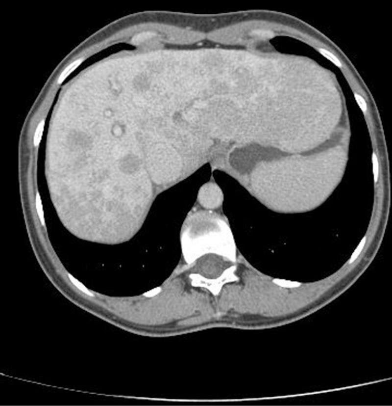
Clinical scenario: A 39-year-old female presented with a CT scan report, that read, ” multiple hypodense lesions in the liver suggestive of metastasis liver. The gall bladder, Hepatic veins, portal vein are normal.     No lesion in the CBD and intrahepatic ducts are also normal”. The patient was admitted for evaluation of the primary after explaining poor prognosis to her husband thoroughly. On examination, she was conscious, oriented and deeply Jaundiced. Her vitals were stable. Her systemic examination was unremarkable.

 Laboratory tests showed normal hemogram but ESR (erythrocyte sedimentation rate )was raised at 52 mm/H (normal, 0-20 mm/H). She had an elevated INR 2.7 Her liver function tests revealed predominantly conjugated hyperbilirubinemia [Bilirubin 12 mg/dl )] elevated liver enzymes (AST aspartate aminotransferase: 818U/L and ALT 51 5 U/L). Serum alkaline phosphatase levels were also elevated at 327 IU/L /L  All her viral markers were negative.

Fig 1 CT scan showing Metastasis Liver

All tumor markers CA -125, CEA and Alpha fetoproteins were elevated. For the search of the primary an upper GI endoscopy, colonoscopy including a mamogram was carried out. All turned out to be normal. While the search for the primary was on , day 4th of admission the patient was found to be incoherent, talking irrelevantly. Her arterial ammonia levels were high and she was shifted to ICU and managed as hepatic encephalopathy. Gladly , her sensorium improved after 3 days and a liver biopsy was planned for evaluation of liver lesions. Keeping in view her high INR 4 units of FFP were transfused and transjugular liver biopsy was done.The Liver biopsy was suggestive of Autoimmune hepatitis a great surprise to one and all.

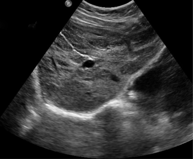
She was given oral steroids and her Liver function tests improved. Repeat Ultrasound of the abdomen showed clearance of liver lesions 

Fig.2 USG abdomen showing no liver lesion post treatment

One year later she was the mother of her 3rd baby.

Take-Home message 

Focal liver lesions are more often discovered with the widespread use of diagnostic imaging modalities. Despite tremendous advancements in the field of radiology, radiological features are not definite. Treat the patient and not an investigation report.

STAGGERING WAITER !

Clinical scenario 

A 57-year-old male patient, waiter by occupation without any previous comorbidities,

came with complaints of the frontal headache of one-week duration

It was associated with, low-grade fever & chills.

The patient also complained of swaying to the left side while walking

which affected his job in the hotel.

There were no similar complaints in the past, no history of prior trauma, head injury, fall or recent vaccination.No history of seizures.No history of loss of weight or appetite. 

On examination: The patient’s vitals and general physical examination were normal.

CNS examination revealed normal higher mental function.

The cranial nerves, motor system, sensory, examination were normal.

The fundus examination was also normal.

He had no signs of meningeal irritation.

However, there was the presence of bilateral cerebellar signs in the form of ataxia,

dysdiadochokinesia, finger nose ataxia more on the left side than the right.

The patient had classical cerebellar gait, 

Other system examination was normal.

Evaluation

On evaluation, the patient had normal Complete blood count  

Hb- 14g%, TLC-8000cells/mm3, platelets, ESR eosinophils we’re in a normal range. 

RBS, LFT, RFT were normal. The serology for  HIV, HBsAg was negative.

MRI brain revealed multifocal ring-enhancing lesions in supra and infratentorial neuroparenchyma with diffuse cerebral edema suggestive of neurocysticercosis.

(Fig 1) 

CSF fluid analysis showed protein 17 mg%, glucose 91mg%, cell count of 18  with neutrophil predominance. CSF ADA was within normal limits.

The ZN stain of CSF was negative for AFB.

Hospital course:

His GCS at presentation was 15. The patient was Treated with anti-edema measures   Mannitol, dexamethasone 8 mg thrice a day.

However during the course of hospital stay patient had a drop in GCS to E4M5V4, 

CT brain was done showed obstructive hydrocephalus and a ventriculoperitoneal shunt

was placed to decompress the brain.

On Post-op day 3 patient’s sensorium improved  and

he was discharged home a few days later

Teaching message 

Bilateral cerebellar signs are more common with systemic diseases

like post-infectious cerebellitis, multiple sclerosis, ADEM.

But in tropical areas, parasitic infestation should also be considered

as a differential for acute ataxia.

Fig2 Neurocystiscircosis in cerebellar region .

\Acute ataxia is an uncommon presentation of neurocysticercosis.

Click to read further  

Diagnosis and Treatment of Neurocysticercosis

This case was contributed by

Dr.Shruthi S

Asst.Prof,

Dept of General Medicine Yenepoya Medical College Mangalore Karnataka

back to school

Friends : Previously we used to read A For Apple and now

A… Anorexia , anemia , asthenia…. R/O Ca stomach

B….Bloating in young may be IBS R/O…organic cause in elderly C….Courvisors law..in a jaundiced patient if gall bladder is palpable ….. it is usually due to extra hepatic malignancy exceptions double …..impaction of stones in CBD &cystic duct,orientalcolangiohepatitis D…..Diarrhea may be IBS in young but evaluate before you label a person IBS R/O microscopic colitis , celiac disease,

E….ECG Changes ….never forget pancreatitis could be a cause..

F…..Fatigue …R /o gut malignancy , chronic hepatitis ,Autoimmunity or Tuberculosis

G….Glossitis R/O iron ,folate B 12 deficiency

H….Hematochezia ….R/O UGI source

I…..Irish nodule (left axillary lymph node)…R/O ca stomach J….Jaundice…. Always evaluate .viral hepatitis , drugs etc R/O ……Biliary obstruction & auto immune hepatitis, etc

K….Koilonychia…r/o Evaluate cause of iron deficiency , colon malignancy in men

L …Large joint arthritis .R/O Inflammatory bowel disease,hepatitis

M…Melena …always evaluate don’t miss right sided colon growth

N….Nausea…drug induced r/o pregnancy in child bearing female.

O…Oral pigmentation …. Peutz.Jeghers to be ruled out

P…Psoas sign…. positive in appendicitis

Q…Quality of life is disturbed more than any complication in IBSR….

Regurgitation likely reflux , psychogenic ,r/o organic cause.

S…Sister Joseph nodule …r/o internal malignancy

T…Trousseaus sign..migratory thrombophilibitis …r/o ca pancreas U….Uveitis R/O Inflammatory bowel disease

V…Virchow node palpable R/O internal malignancy

W…Water brash…Gastro esophagial reflux

X….Xanthomas ..R/O Lipid abnormalities

Y….Yellow nail patella syndrome

Z…..Zollinger Ellison syndrome….

Disturbed by gas ?

Disturbed by GAS in the bowel ?
The excessive flatulence can be embarrassing and make you feel uncomfortable around others. However, it can usually be controlled with changes to your diet and lifestyle. Flatulence is a normal biological process and is something everyone experiences regularly.
Some people pass wind only a few times a day, others a lot more, but the average is said to be about 5 to 15 times a day.

Continue reading Disturbed by gas ?

Shortness of Breath in a young male

Clinical scenario: A 30-year-old male presented with a history of shortness of breath of one-day duration. He denied a history of fever, PND, orthopnea or palpitations.On examination, the patient was conscious, oriented hemodynamically stable.

Continue reading Shortness of Breath in a young male

A careful examination clinched the diagnosis

Clinical scenario:

A 45-year-old farmer, presented with complaints of fever, chills, generalized weakness associated with yellowish discoloration of sclera for 15 days.

Continue reading A careful examination clinched the diagnosis

OLD MAN WITH FEVER & ALTERED SENSORIUM

Clinical scenario :

A 60-year-old male presented with complaints of easy fatigability of 3 months duration .it was followed by a fever of 3 weeks duration Fever was not associated with Rigors and Chills more so in the evenings. The patient also complained of reduced appetite and weight loss-16kgs in 3 months. Before the current presentation, he developed progressive deterioration in his sensorium and vomiting. The vomiting was not bilious, not blood-tinged usually the food taken by him earlier. There was no history of cough with expectoration, no history of diarrhea, no dysphagia No history of diabetes, hypertension in the past or any other comorbidity

Continue reading OLD MAN WITH FEVER & ALTERED SENSORIUM

Upper gi endoscopy indications

Upper endoscopy is currently one of the most frequently performed procedures. The most common indications for 

Diagnostic EGD include 

Dyspepsia unresponsive to medical therapy or associated with systemic signs, dysphagia or odynophagia, persistent gastroesophageal reflux symptoms, occult gastrointestinal bleeding, and surveillance for malignancy. 

Continue reading Upper gi endoscopy indications

Indications of colonoscopy

Indications for colonoscopy:
1.Lower GI bleeding
2.Screening and surveillance of colorectal polyps and cancers:
a. Colon cancer
b. Surveillance after polypectomy
c. Colorectal cancer post-resection surveillance
d. Inflammatory bowel diseases
3.Acute and chronic diarrhea
4.Therapeutic indications for colonoscopy:
a. Excision and ablation of lesions
b. Treatment of lower GI bleeding
c. Colonic decompressiond. Dilation of colonic stenosise. Foreign body removal
5.Miscellaneous indications:
a. Abnormal radiological examinations.
b. Isolated unexplained abdominal pain
c. Chronic constipation
d. Preoperative and intraoperative localization of colonic lesions
Continue reading Indications of colonoscopy

Use antibiotics wisely

Today about ANTIBIOTIC RESISTANCE;
It’s an emerging issue which will pose the most serious threat to all of us as many bacteria are rapidly becoming resistant to many antibiotics.
The main reason is frequent use of antibiotics without proper indication.

Continue reading Use antibiotics wisely

Knowledge is power!